The visualization and imagination of biological structures at sub-microscopic scales is one of the defining features of modern biology. Our sense of the materiality of the sub-microscopic world depends on the prolific visual culture of the life sciences: for example, Watson and Crick’s telegenic DNA model turned the double helix into a generic symbol of biological essence, while the now-ubiquitous images of the SARS-CoV-2 virion are a constant reminder that the invisible spread of this disease is caused by a material entity. These images are not merely representations of scientific research: our theoretical and intuitive grasp of invisible structures and pathogens has depended on optical technologies and optical methodologies that provide visual evidence of their form.
This project will examine the history of the light microscope, x-ray diffractometer, and the electron microscope as optical technologies that are grounded in constellations of material practices in molecular biology and virology. That is to say, this project will examine how the creation of images of the molecular structure of fibers and viruses relies on manipulation, preparation, and modeling of the submicroscopic objects that are imaged. I use the term “optical culture” in contrast to the art historical term “visual culture,” in order to highlight the material and technical demands of light microscopy, x-ray crystallography, and electron microscopy. These laboratory technologies demanded intimate, if incomplete, knowledge of the specimen to be imaged, as well as knowledge of the optical possibilities (and limitations) of their instruments. Therefore, the project will investigate the material practices that lay behind micrograms and x-ray diagrams—images which worked to affirm the material reality of the invisible entities being studied.
The project examines three historical case studies. The first is a multi-sited examination of theories of fiber structure in the 1920s and 1930s which were based on x-ray, light microscopical, and viscometrical studies of leather, wool, and linen. The second case study examines work by the virologist June Almeida (1930–2007), who identified the family of coronaviruses by electron microscopy in 1966–1967. This case study will examine Almeida’s pathbreaking methods of virus taxonomy, including culturing viruses in vitro, developing biochemical methods to aggregate viruses for imaging, and creating models of how viruses might hypothetically interact with the electron beam (see image below). The third case study examines the race to discover the cause of the AIDS pandemic in 1982–1984, and the role electron microscopy played in disputes over the identification and taxonomy of LAV/HTLV-III/ARV, later renamed HIV-1.
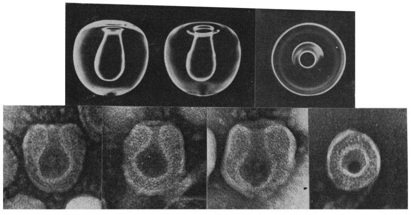
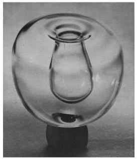
A glass model (above) used to study the internal structure of prepared coronavirus (avian infectious bronchitis virus) treated with formaldehyde. R. W. Bingham and June D. Almeida, “Studies on the Structure of a Coronavirus: Avian Infectious Bronchitis Virus,” Journal of General Virology 36, no. 3 (Sept. 1977): 495–502. doi:10.1099/0022-1317-36-3-495.